 We use the iCare Tonometer!
For easy, accurate and patient-friendly intra-ocular pressure measurement.
iCare tonometers are based on unique, patented rebound technology, in which a very light and small probe is used to make a momentary contact with the cornea. No specialized skills for its use the quick and painless measurement is barely noticed by the patient and and any anesthesia or inconvenient air puffs are not needed at all.Icare technology has been proven accurate and reliable by several clinical studies which demonstrates the same accuracy and uncompromised reliability in its measurements as the Golden standard i.e. complies to ISO 8612 tonometer standard, and better results than other hand-held tonometers presently on the market. Never before has intra-ocular pressure (IOP) measuring been this easy – and still accurate!
No Dilation Drops Or Air Puffs
No more blurry eyes! Our technology eliminates the need for dilation drops in most cases. This means you can resume your activities as soon as you leave our facility with healthy eyes and clear vision.
Corneal Mapping
Corneal topography, also known as photokeratoscopy or videokeratography, is a non-invasive medical imaging technique for mapping the surface curvature of the cornea, the outer structure of the eye. Since the cornea is normally responsible for some 70% of the eye’s refractive power, its topography is of critical importance in determining the quality of vision.
The three-dimensional map is therefore a valuable aid to the examining ophthalmologist or optometrist and can assist in the diagnosis and treatment of a number of conditions; in planning refractive surgery such as LASIK and evaluation of its results; or in assessing the fit of contact lenses. A development of keratoscopy, corneal topography extends the measurement range from the four points a few millimeters apart that is offered by keratometry to a grid of thousands of points covering the entire cornea. The procedure is carried out in seconds and is completely painless.
OCT Scans
We use cutting-edge digital imaging technology to assess your eyes. Many eye diseases, if detected at an early stage, can be treated successfully without total loss of vision. Your retinal Images will be stored electronically. This gives the eye doctor a permanent record of the condition and state of your retina.
This is very important in assisting your Fort Lauderdale optometrist to detect and measure any changes to your retina each time you get your eyes examined, as many eye conditions, such as glaucoma, diabetic retinopathy and macular degeneration are diagnosed by detecting changes over time.
The advantages of digital imaging include:
- Quick, safe, non-invasive and painless
- Provides detailed images of your retina and sub-surface of your eyes
- Provides instant, direct imaging of the form and structure of eye tissue
- Image resolution is extremely high quality
- Uses eye-safe near-infra-red light
- No patient prep required

iVue OCT by Optovue
We are proud to offer the Optovue- iVue OCT. The iVue is a comprehensive OCT platform that allows us to have retinal, optic nerve and anterior segment imaging capabilities, as well as the exclusive iWellness scan. This quick and easy scan provides an in-depth look at the health of your retina and other portions of the inner eye, which helps us identify signs of disease before they would be noticed on an eye exam which did not include this in-depth eye scan.
An OCT scan is a noninvasive, painless test. It is performed in about 10 minutes right in our office. Feel free to contact Fidler Eye Care to inquire about an OCT at your next appointment.
Digital Retinal Imaging
Digital Retinal Imaging allows your eye doctor to evaluate the health of the back of your eye, the retina. It is critical to confirm the health of the retina, optic nerve and other retinal structures. The digital camera snaps a high-resolution digital picture of your retina. This picture clearly shows the health of your eyes and is used as a baseline to track any changes in your eyes in future eye examinations.
We use our enhance digital imaging to give you the best care possible.
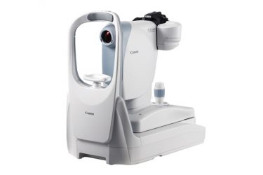 Canon Fundus Digital Imaging
Ocular fundus imaging plays a key role in monitoring the health status of the human eye. Currently, a large number of imaging modalities allow the assessment and/or quantification of ocular changes from a healthy status. Features the latest in Canon retinal imaging technology and enhancements in a compact and lightweight design. Fundus imaging is essential in the diagnosis and monitoring of various ophthalmic diseases. A major advantage is that the doctor can share these images with specialists at other locations and/or times, they can also magnify areas of interest that cannot be easily seen with a handheld ophthalmoscope.
Optical Coherence Tomography (OCT)
An Optical Coherence Tomography scan (commonly referred to as an OCT scan) is the latest advancement in imaging technology. Similar to ultrasound, this diagnostic technique employs light rather than sound waves to achieve higher resolution pictures of the structural layers of the back of the eye.
A scanning laser used to analyze the layers of the retina and optic nerve for any signs of eye disease, similar to an CT scan of the eye. It works using light without radiation, and is essential for early diagnosis of glaucoma, macular degeneration and diabetic retinal disease.
With an OCT scan, doctors are provided with color-coded, cross-sectional images of the retina. These detailed images are revolutionizing early detection and treatment of eye conditions such as wet and dry age-related macular degeneration, glaucoma, retinal detachment and diabetic retinopathy.
An OCT scan is a noninvasive, painless test. It is performed in about 10 minutes right in our office. Feel free to contact our office to inquire about an OCT at your next appointment.
Visual Field Testing
A visual field test measures the range of your peripheral or “side” vision to assess whether you have any blind spots (scotomas), peripheral vision loss or visual field abnormalities. It is a straightforward and painless test that does not involve eye drops but does involve the patient’s ability to understand and follow instructions.
Although the test can be used to detect defects caused by various medical conditions such as stroke, brain tumors or other neurological deficits, it is most frequently used in eye care practices to diagnose, monitor or rule out glaucoma.
 Humphrey Zeiss Visual Field Analyzer
The visual field test is carried out by a computerized machine, we use the state of the art Humphrey™ Field Analyzer. This new Visual field testing provides fast supra-threshold testing complete with statistical analysis in about 4 minutes. Visual field testing is a basic element in the standard care for glaucoma and all neurological vision loss. We are proud to screen everyone in our pre-testing with this proven diagnostic performance in detecting early visual field loss.
The test primarily measures the quality and quantity of peripheral or “side” vision you have. It is a straightforward and painless test which does not involve eye drops. Essentially, lights are flashed on and you have to press a button whenever you see the light. Your head is kept still on a chin rest. The lights are bright or dim at different stages of the test. Some of the flashes are to check if you are concentrating. Each eye is tested separately and the entire test takes less than 20 minutes.
Visit Fidler Eye Care for Computer Eyestrain evaluation and consultation.
Zeiss Clarus 500
Color. Clarity. Comfort.
The advent of widefield retinal imaging has shown us that indications of disease are often located in the far periphery of the retina. CLARUS TM 500 is the next generation fundus imaging system from ZEISS that provides true color and high-resolution across an entire ultra-widefield image.
iWellness OCT scan with the iVue OCT by Optovue
We are proud to offer the Optovue- iVue OCT. The iVue is a comprehensive OCT platform that allows us to have retinal, optic nerve and anterior segment imaging capabilities, as well as the exclusive iWellness scan. This quick and easy scan provides an in-depth look at the health of your retina and other portions of the inner eye, which helps us identify signs of disease before they would be noticed on an eye exam which did not include this in-depth eye scan.